


We Emit Visible Light That Fades When We Die, and Scientists Captured It on Camera

A team of Canadian researchers placed four mice in a dark box and pointed ultra sensitive cameras at them. The cameras detected something the human eye cannot see: individual particles of light emerging from the animals’ living tissue.
The researchers then euthanized the mice and kept imaging. The light did not stop entirely, but its intensity dropped measurably. Something that had been present while the animals lived had diminished after death.
The findings, published in a physical chemistry journal, add to a body of research on ultraweak biological emissions that has slowly accumulated over decades. They also invite comparison to earlier photographic techniques that attracted paranormal interpretations before being debunked.
The Body’s Faint Glow Before and After Death
A team from the University of Calgary and the National Research Council of Canada documented a distinct decrease in visible light emanating from living mice after the animals were euthanized, according to findings published in The Journal of Physical Chemistry Letters.

The researchers used electron multiplying charge coupled device cameras to image four immobilized mice for one hour while alive, then for another hour after euthanasia. The mice were warmed to maintain body temperature throughout the second imaging period to eliminate heat as a variable. The cameras detected individual photons in the visible band emerging from cells both before and after death, with a statistically significant reduction in the post euthanasia measurements.
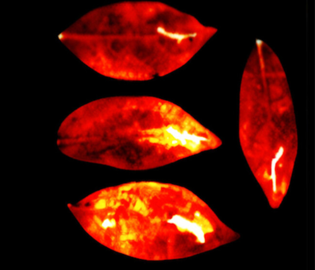
Similar results were obtained from thale cress and dwarf umbrella tree leaves subjected to physical injury and chemical agents. The researchers wrote in their paper that “the injury parts in all leaves were significantly brighter than the uninjured parts of the leaves during all 16 hours of imaging.”
The phenomenon, described in the paper as ultraweak photon emission, has been documented in isolated biological materials for decades. Cow heart tissue and bacterial colonies are among specimens previously shown to produce spontaneous light in the 200 to 1,000 nanometer range, as detailed in research published in MicrobiologyOpen. The Calgary led experiment extended those observations to whole animal subjects.
Electron multiplying CCD cameras, first introduced in 2001, enable detection of single photon events through an on chip multiplication structure that amplifies charge signals before readout. This architecture bypasses the readout noise that limits sensitivity in conventional CCD and CMOS sensors, according to Oxford Instruments. The technology allows imaging of light intensities far below what standard cameras can register.
The study’s authors propose that reactive oxygen species are the primary source of the emissions. Living cells produce these molecules when subjected to stressors including heat, toxins, pathogens or nutrient deprivation. Hydrogen peroxide and similar compounds can trigger transformations in fats and proteins that elevate electrons to higher energy states; photons are released when electrons return to baseline positions.
Visible light occupies the 380 to 700 nanometer range of the electromagnetic spectrum, according to NASA. The human eye detects this band through cone shaped retinal cells, but the photon flux documented in the Calgary experiment falls below the threshold of unassisted human vision.
What Kirlian Photography Got Wrong
The technique has precedents in earlier imaging methods that attracted paranormal interpretations. Kirlian photography, developed in 1939 by Soviet engineer Semyon Kirlian, produces images of coronal discharges when objects on photographic plates are connected to high voltage sources. The method gained attention in parapsychology circles during the 1970s following claims that the resulting images depicted life fields or auras reflecting emotional and physical states.

Those claims were rejected by the scientific community. Research published in 1973 in the Journal of Applied Physics demonstrated that variations in corona discharge streamer length, density and color correlate with surface moisture content rather than any hypothesized biological energy field. Later experiments showed that cleaning imaging surfaces eliminates the “phantom leaf” effect sometimes presented as evidence of persistent energy fields after tissue removal.
The Calgary led study did not use high voltage discharge techniques. The authors explicitly frame their work as a biophoton investigation rather than an attempt to validate earlier Kirlian claims, a distinction noted in ScienceAlert’s reporting on the findings. They describe potential applications in non invasive monitoring of tissue stress in human patients, agricultural crops or bacterial samples.
Why Clinical Use Remains Years Away
The study’s limitations include the small sample size of four mice and the controlled laboratory conditions required for imaging. Ambient light overwhelms the ultraweak emissions, necessitating dark box confinement of subjects. Whether the technique can be adapted for clinical use in humans remains untested.
The researchers reported that the emissions remained detectable but significantly reduced after death, suggesting the photon production depends on ongoing metabolic processes rather than passive physical properties of tissue. Reactive oxygen species generation ceases when cellular respiration stops, which is consistent with the observed post mortem decline.
The National Research Council of Canada and the University of Calgary have not announced follow up studies or clinical trials as of today. The paper states that remote monitoring of stress through ultraweak photon detection “could provide technicians and medical specialists with a powerful, non invasive research or diagnostics tool,” but does not specify a timeline for translational work.
Enjoyed this article? Subscribe to our free newsletter for engaging stories, exclusive content, and the latest news.
What's Your Reaction?
 Like
0
Like
0
 Dislike
0
Dislike
0
 Love
0
Love
0
 Funny
0
Funny
0
 Angry
0
Angry
0
 Sad
0
Sad
0
 Wow
0
Wow
0